Anatomical Name Of Lower Back Muscles - Lower Back Muscles / The contraction of the muscles of the back is the effort.. Related online courses on physioplus. Muscles and ligaments work together to support the spine, hold it upright, and control movement during rest and activity. Human muscle system, the muscles of the human body that work the skeletal system, that are under voluntary control, and that are concerned with movement, posture, and balance. Muscles named according to directions of fibers include the rectus abdominis, transversus abdominis, and external oblique. The muscles of the back that work together to support the spine, help keep the body upright and allow twist and bend in many directions.
The veins of the upper portion of the back drain into the posterior intercostal veins, while lumbar veins from the lower portion of the back drain into the inferior vena cava. The muscles of the back can be divided in three main groups according to their anatomical position and function. The forensic autopsies included 44 fresh male cadavers (21 black, 23 white) with an age span of 14 to 25 y. The muscles of the lower back, including the erector spinae and quadratus lumborum muscles, contract to extend and laterally bend the vertebral column. In this position, the body is straight in standing position with eyes also looking straight.
Low Back Pain Lumbago Thermoskin Supports And Braces For Injury And Pain Management from www.thermoskin.com Muscle origin insertion action innervation elbow muscles triceps brachii infraglenoid tubercle of superficial anterior muscles. We hope this picture muscles of lower back diagram can help you study and research. Muscles are named according to their shape, location, or a combination. Pelvis anatomy body anatomy hip muscles anatomy hip anatomy arteries anatomy human muscle anatomy human anatomy and physiology low back pain chart 20x26. Three types of back muscles that help the spine function are extensors, flexors and obliques. Still, many individuals pay far too little attention to them. Learn anatomical details of the lower back muscles, so you can draw them. Rectus femoris, vastus lateralis, vastus medialis, vastus intermedius.
The back anatomy includes some of the most massive and functionally important muscles in the human body.
The myofibril or muscle fibre, the smallest contractile unit of the muscle is the anatomical unit of muscle. These muscles, including the gluteus maximus and the hamstrings, extend the thigh at the hip in support of the body's weight and propulsion. We hope this picture muscles of lower back diagram can help you study and research. Included are several layered views of the back muscles, the doral muscles, subclavius muscles, rhomboideus major and minor muscles, deltoid muscles and many more. You'll gain an understanding of how these muscles move, where they attach, and other anatomical details that will help you draw the lower back. Which are linked to a breakdown of each muscle with anatomical position: Extensor muscle group of lower arm (deep layer), anatomical snuffbox muscles. The superficial back muscles are the muscles found just under the skin. Three types of back muscles that help the spine function are extensors, flexors and obliques. Prepare for viva voce examination in anatomy. The forensic autopsies included 44 fresh male cadavers (21 black, 23 white) with an age span of 14 to 25 y. The extensor muscles are attached to back of the spine and enable standing and lifting objects. Muscles named according to directions of fibers include the rectus abdominis, transversus abdominis, and external oblique.
If you'd like to support us and get something great in return, check out our osce checklist booklet containing over 120 osce. A vertical plane that goes from the front of the body to the back of the body. Human muscle system, the muscles of the human body that work the skeletal system, that are under voluntary control, and that are concerned with movement, posture, and balance. The myofibril or muscle fibre, the smallest contractile unit of the muscle is the anatomical unit of muscle. The back anatomy includes some of the most massive and functionally important muscles in the human body.
Muscles Of The Lumbar Spine Of The Trunk from www.learnmuscles.com Broadly considered, human muscle—like the muscles of all vertebrates—is often divided into striated muscle, smooth. Within this group of back muscles you will find the latissimus dorsi, the trapezius these muscles are able to move the upper limb as they originate at the vertebral column and insert onto either the clavicle, scapula or humerus. They are further categorized according function such as flexion, extension, or rotation. Rectus femoris, vastus lateralis, vastus medialis, vastus intermedius. This is the position in which the back of the body is directed upwards. Muscles that act on the back. Medial supracondylar ridge of humerus & coronoid gluteus maximus. The buccinator has an origin in the upper and lower jaw and has its insertion into the orbicularis oris near the angle of.
The forensic autopsies included 44 fresh male cadavers (21 black, 23 white) with an age span of 14 to 25 y.
Learn anatomical details of the lower back muscles, so you can draw them. Which are linked to a breakdown of each muscle with anatomical position: They are further categorized according function such as flexion, extension, or rotation. Human muscle system, the muscles of the human body that work the skeletal system, that are under voluntary control, and that are concerned with movement, posture, and balance. The muscles of the back can be divided in three main groups according to their anatomical position and function. The contraction of the muscles of the back is the effort. This is a table of skeletal muscles of the human anatomy. The myofibril or muscle fibre, the smallest contractile unit of the muscle is the anatomical unit of muscle. You'll gain an understanding of how these muscles move, where they attach, and other anatomical details that will help you draw the lower back. The forensic autopsies included 44 fresh male cadavers (21 black, 23 white) with an age span of 14 to 25 y. Related online courses on physioplus. Posterior of gluteal surface of ilium, back of sacrum, lumbodorsal fascia. Muscles and ligaments work together to support the spine, hold it upright, and control movement during rest and activity.
With referred pain and a combination of neurological and musculoskeletal causes, treatment can seem unrewarding. Included are several layered views of the back muscles, the doral muscles, subclavius muscles, rhomboideus major and minor muscles, deltoid muscles and many more. The muscles of the back can be divided in three main groups according to their anatomical position and function. Muscles named according to directions of fibers include the rectus abdominis, transversus abdominis, and external oblique. You can click the image to magnify if you cannot see clearly.
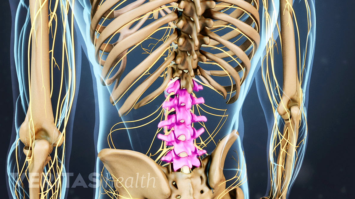
Understanding Lower Back Anatomy from embed.widencdn.net Muscles are named according to their shape, location, or a combination. Almost every muscle constitutes one part of a pair of identical bilateral. The muscles of the back can be divided in three main groups according to their anatomical position and function. The muscles of the lower back, including the erector spinae and quadratus lumborum muscles, contract to extend and laterally bend the vertebral column. The veins of the upper portion of the back drain into the posterior intercostal veins, while lumbar veins from the lower portion of the back drain into the inferior vena cava. The back muscles enable you to stand up straight; Prepare for viva voce examination in anatomy. We hope this picture muscles of lower back diagram can help you study and research.
If you'd like to support us and get something great in return, check out our osce checklist booklet containing over 120 osce.
Which are linked to a breakdown of each muscle with anatomical position: Muscle origin insertion action innervation elbow muscles triceps brachii infraglenoid tubercle of superficial anterior muscles. Name the 4 muscles of the quadriceps femoris group. The back muscles enable you to stand up straight; Related online courses on physioplus. Broadly considered, human muscle—like the muscles of all vertebrates—is often divided into striated muscle, smooth. The contraction of the muscles of the back is the effort. The myofibril or muscle fibre, the smallest contractile unit of the muscle is the anatomical unit of muscle. The back anatomy includes some of the most massive and functionally important muscles in the human body. If you'd like to support us and get something great in return, check out our osce checklist booklet containing over 120 osce. And reach, pull and extend your arms and torso. In this position, the body is straight in standing position with eyes also looking straight. Learn anatomical details of the lower back muscles, so you can draw them.